Home » Without Label » 48+ neu Sammlung Diagram Inner Ear : Diagram Of A Human Ear Vector Illustration Of Diagram Of ... / Nervous system disease nerve diagram human body, others, hand, symmetry, head png.
48+ neu Sammlung Diagram Inner Ear : Diagram Of A Human Ear Vector Illustration Of Diagram Of ... / Nervous system disease nerve diagram human body, others, hand, symmetry, head png.
48+ neu Sammlung Diagram Inner Ear : Diagram Of A Human Ear Vector Illustration Of Diagram Of ... / Nervous system disease nerve diagram human body, others, hand, symmetry, head png.. The bony labyrinth, a cavity in the temporal bone, is divided into three sections: (iv) the middle ear is connected with the inner ear through two small openings closed by the membranes. The inner ear houses the sensory organs that help in hearing and maintaining balance. Diagram showing the structure of the human ear, detailing the parts of the outer, middle, and inner ear. In mammals, it consists of the bony labyrinth.
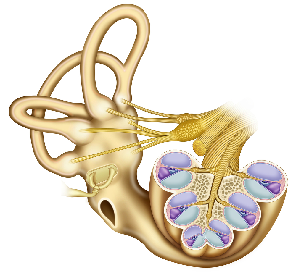
The inner ear consist of the cochlea, the balance mechanism, the vestibular and auditory nerve. The inner ear is found in all vertebrates, with substantial variations in form and function. Sensory structures within the vestibule and semicircular canals control this. In vertebrates, the inner ear is mainly responsible for sound detection and balance. It is lined with special sensory cells called hair cells which are sensitive to sound.
Hearing: Sensing the World Through Sound Waves from www.medicalsciencenavigator.com Inner ear malformations are a spectrum of congenital anomalies involving the inner ear structures with an emphasis on the cochlea due to their implications for sensorineural hearing loss. Learn vocabulary, terms and more with flashcards, games and other study tools. In vertebrates, the inner ear is mainly responsible for sound detection and balance. However, you may skip the diagram of the inner ear as it is never asked. Nervous system disease nerve diagram human body, others, hand, symmetry, head png. The inner ear houses the sensory organs that help in hearing and maintaining balance. You can see in the diagram that the external ear captures the traveling sound waves. A framework for speechreading acquisition tools.
However, you may skip the diagram of the inner ear as it is never asked.
Military disability ratings for ear conditions. It is lined with special sensory cells called hair cells which are sensitive to sound. Sensory structures within the vestibule and semicircular canals control this. The bony labyrinth, a cavity in the temporal bone, is divided into three sections: The inner ear is also responsible for helping maintain balance. You can see in the diagram that the external ear captures the traveling sound waves. The inner ear is found in all vertebrates, with substantial variations in form and function. In mammals, it consists of the bony labyrinth. Learn vocabulary, terms and more with flashcards, games and other study tools. The inner ear is one of the most complex component of the described entity. Structure and anatomy the anatomy of ear consists of external ear, middle ear and inner ear. These openings are (a) fenestra ovalis (oval window) as mentioned above and (b). The inner ear (internal ear, auris interna) is the innermost part of the vertebrate ear.
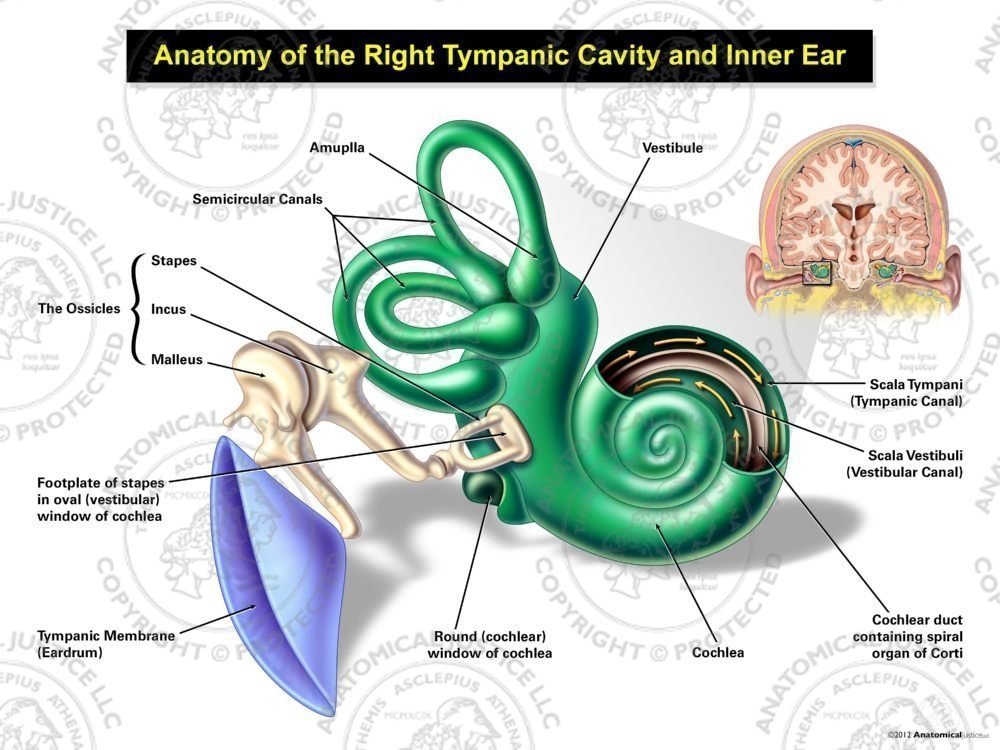
Anatomy of the Right Tympanic Cavity and Inner Ear from anatomicaljustice.com The inner ear is separated from the brain space filled with a viscous fluid. Nervous system disease nerve diagram human body, others, hand, symmetry, head png. The inner ear (internal ear, auris interna) is the innermost part of the vertebrate ear. The inner ear is one of the most complex component of the described entity. The vestibule contains two sacs, the utricle and the saccule. Structure and anatomy the anatomy of ear consists of external ear, middle ear and inner ear. She is responsible for conducting sounds. The bony labyrinth, a cavity in the temporal bone, is divided into three sections:
The inner ear is found in all vertebrates, with substantial variations in form and function.
You can see in the diagram that the external ear captures the traveling sound waves. There is a printable worksheet available for download here so you can take the quiz with pen and paper. There are actually two labyrinths of the inner ear, one inside the other, the membranous labyrinth contained within the bony labyrinth. .labels | inner ear diagram we provide you here simple ear diagram in easy way for drawing.also eardrum (c) a hole in the eardrum #hearingloss #conductivehearingloss #sound #outerear #innerear. Inner ear malformations are a spectrum of congenital anomalies involving the inner ear structures with an emphasis on the cochlea due to their implications for sensorineural hearing loss.
Shut Up, Chris Brown! - SiOWfa12: Science in Our World ... from www.personal.psu.edu The inner ear is found in all vertebrates, with substantial variations in form and function. The vestibule contains two sacs, the utricle and the saccule. Inner ear diagram labeled inner ear diagram labeled inner. The inner ear is one of the most complex component of the described entity. Inner ear diagram 9 mammaliaform inner ear evolution cochlear canal curvature and. Sensory structures within the vestibule and semicircular canals control this. Download scientific diagram | 1: (iv) the middle ear is connected with the inner ear through two small openings closed by the membranes.
Diagram showing the structure of the human ear, detailing the parts of the outer, middle, and inner ear.
In this article, you will learn more about the inner ear's anatomy. Inner ear malformations are a spectrum of congenital anomalies involving the inner ear structures with an emphasis on the cochlea due to their implications for sensorineural hearing loss. Inner ear diagram labeled inner ear diagram labeled inner. These openings are (a) fenestra ovalis (oval window) as mentioned above and (b). The vestibule contains two sacs, the utricle and the saccule. Sensory structures within the vestibule and semicircular canals control this. The inner ear consist of the cochlea, the balance mechanism, the vestibular and auditory nerve. Copyright • • twp parts of inner ear. The inner ear houses the sensory organs that help in hearing and maintaining balance. Anatomy of the inner ear by annie campbell. The diagrams of the eye (there are two, so both of them) are very important and can be asked. The inner ear is separated from the brain space filled with a viscous fluid. The inner ear (internal ear, auris interna) is the innermost part of the vertebrate ear.